Articolo del 05/05/2025
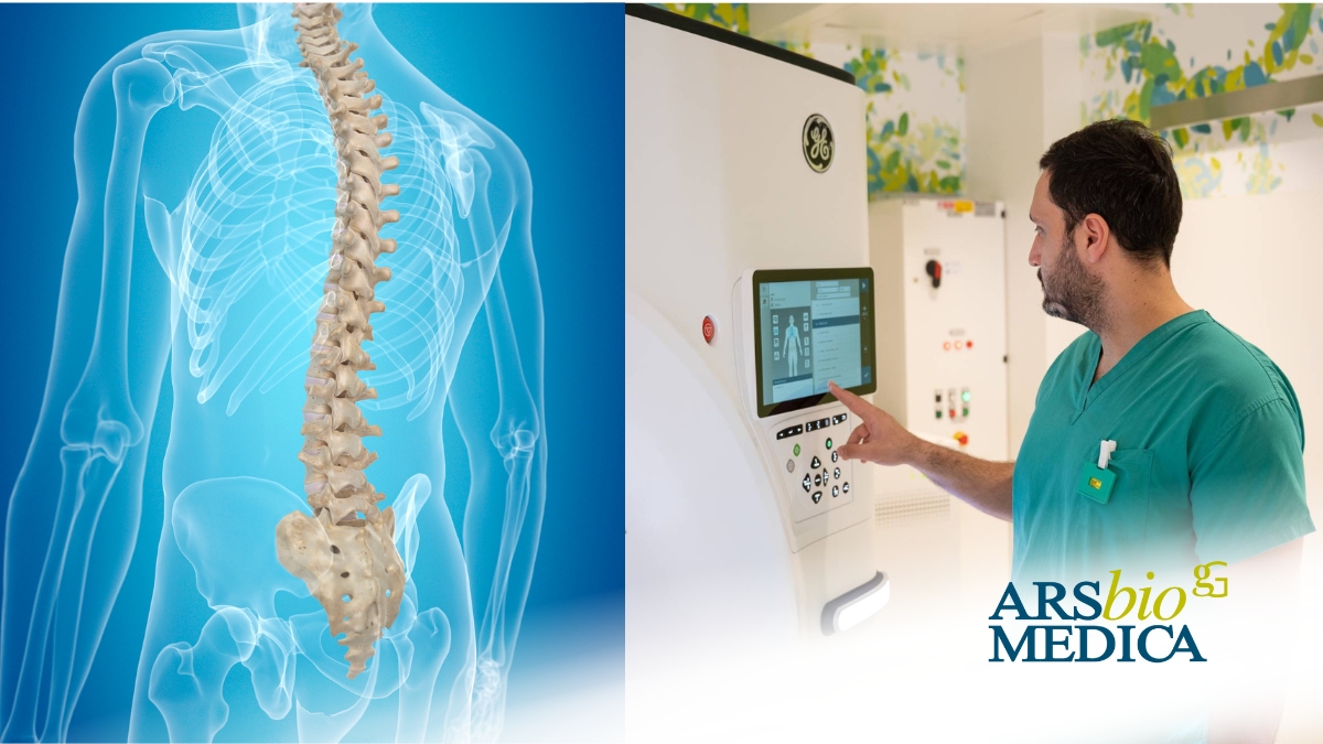
A CT scan of the spine is a diagnostic imaging test that uses ionizing radiation to produce highly detailed images of the vertebrae, intervertebral discs, spinal canal, and surrounding structures. It allows for the rapid detection and assessment of fractures, degenerative changes, inflammatory conditions, and neoplastic lesions.
What are the main clinical indications for this examination?
And why is it advantageous to undergo this scan at Arsbiomedica using a 512-slice CT scanner?
We explore the topic with Dr. Nicolò Rumi, radiologist at Arsbiomedica.
The most common indications include persistent pain, major trauma with suspected fractures, complex scoliosis, infections, tumors, and pre-operative assessments.
Arsbiomedica’s 512-slice CT scanner provides ultra-high-definition images within seconds, ensuring greater diagnostic precision while minimizing radiation exposure. Additionally, the dual-energy technology significantly reduces beam-hardening artifacts from metallic implants in post-surgical patients and enables the identification of bone marrow edema in cases where MRI is contraindicated.
What are the advantages of 3D reconstruction compared to traditional 2D imaging?
Compared to conventional 2D imaging, 3D reconstruction offers volumetric visualization of the spine from every angle. This allows for a more comprehensive understanding of complex lesions, enhances the accuracy of surgical planning, and facilitates clearer communication of the clinical picture to both patients and fellow specialists.
Are there any limitations or contraindications to using 3D CT reconstruction?
The main limitations are related to the quality of the initial images—patient movement or the presence of metallic devices can compromise reconstruction accuracy. However, our 512-slice dual-energy CT scanner overcomes many of these challenges through rapid acquisition and a dedicated MAR (Metal Artifact Reduction) system.
There are no contraindications to performing 3D reconstruction.
Can you share a practical example where 3D CT reconstruction made a difference in diagnosis or treatment?
In one case, high-resolution CT imaging with MAR and 3D reconstruction enabled the detailed evaluation of a complex post-traumatic spinal fracture in a patient with a previous spinal surgery. This allowed the surgeon to precisely plan the procedure, reducing operative time and significantly improving the patient’s functional outcome.


